Digital smile design has shifted aesthetic dentistry from subjective craftsmanship toward objective, data-driven planning. Patients seeking cosmetic enhancements—whether clear aligners, porcelain veneers, or full-mouth restorations—expect predictable outcomes that harmonize with their unique facial features. Achieving this standard requires meticulous data collection from the initial consultation through final restoration placement.
At the center of this transformation is the dental 3D scanner. By replacing traditional elastomeric impression materials with high-definition optical data, clinicians and dental laboratories can analyze, design, and execute aesthetic treatment plans with unprecedented clinical accuracy. Integrating a modern intraoral scanner into the diagnostic workflow streamlines patient communication, enhances interdisciplinary collaboration, and ensures long-term restorative fit.
- Capturing High-Fidelity Diagnostic Data at the Chairside
A successful smile design relies on the accuracy of initial diagnostic models. Traditional impression techniques using alginate or polyvinyl siloxane (PVS) present inherent challenges. Material shrinkage, micro-voids, tray flexure, and distortion during transport to the dental laboratory can compromise model fidelity. Furthermore, physical impressions are uncomfortable for patients and frequently require time-consuming retakes.
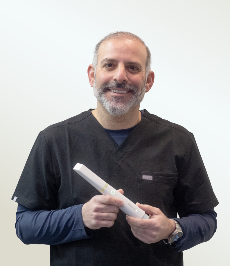
Using a modern intraoral scanner eliminates these variables by capturing hard and soft tissue geometry directly within the oral cavity. High-resolution optical sensors record surface contours, interproximal contact zones, and gingival margins in real time.
Direct Digital Impressions: Scanning generates accurate 3D surface files (such as STL, PLY, or OBJ formats) without material degradation.
Immediate Quality Control: Clinicians can inspect scan data on screen instantly, verifying margin clarity, preparation clearance, and bite registration before the patient leaves the chair.
Enhanced Comfort: Non-invasive scanning improves the patient experience, making diagnostic data collection fast and efficient.
- Harmonizing Intraoral Scans with Facial Aesthetics
Smile design extends beyond isolated dental arches; it requires analyzing how teeth frame the lips, face, and dynamic expressions. A static 2D photograph provides basic visual guidance, but it lacks spatial depth and cannot accurately simulate dynamic jaw movement or multi-angle lighting.
Combining an intraoral scanner dataset with 3D facial scanning technology creates a comprehensive digital patient model. Within the SHINING 3D DENTAL software ecosystem, clinicians can merge intraoral scans captured by devices like the Aoralscan series with 3D facial scans (such as MetiSmile data).
This multi-layered digital alignment enables clinicians to analyze key aesthetic parameters in three dimensions:
Facial Midline Alignment: Positioning central incisors precisely relative to the facial axis.
Incisa-Lip Dynamics: Evaluating tooth display at rest and during full smile movement.
Occlusal Plane Orientation: Aligning the interpupillary line with the occlusal plane to prevent tilted aesthetic outcomes.
By overlaying high-accuracy intraoral surfaces onto 3D facial structures, dental teams can design restorations that naturally complement the patient’s overall facial geometry.
- Increasing Clinical Predictability for Complex Restorations
In restorative and cosmetic dentistry, subtle margin errors can result in micro-leakage, unnatural contours, or premature restoration failure. A high-performance dental 3D scanner provides the spatial resolution necessary to plan complex aesthetic cases, including multi-unit veneers, crown preparations, and full-arch rehabilitations.
Advanced scanning hardware utilizes structured light or intraoral photogrammetry (IPG) technology to capture surface topology with exceptional accuracy. Systems developed by SHINING 3D DENTAL maintain consistent accuracy across full-arch scans, minimizing spatial distortion across the dental arch.
This structural accuracy improves several clinical steps:
Margin Definition: Clear digital display of crown and veneer prep margins facilitates precise CAD margin marking.
Dynamic Occlusion Analysis: Capturing real-time bite relationships allows technicians to design functional occlusion that prevents premature contacts on aesthetic restorations.
Diagnostic Mock-ups: Scanned data serves as the foundation for digital wax-ups, allowing clinicians to print physical mock-ups for chairside intraoral try-ins prior to tooth preparation.
- Enhancing Patient Engagement and Treatment Acceptance
Visual communication is essential during aesthetic treatment planning. Patients often struggle to visualize proposed treatment outcomes based solely on verbal descriptions or 2D photo edits. A 3D digital approach transforms the consultation process into an interactive visual experience.
When clinicians present a 3D model generated by a dental 3D scanner, patients can view their current dentition from any angle alongside proposed digital smile simulations.
Interactive Consultations: Dentists can manipulate 3D models chairside, pointing out structural wear, alignment issues, or gingival asymmetry.
Realistic Simulations: Software tools allow real-time adjustments to tooth shape, shade, and length, allowing patients to participate directly in their aesthetic choices.
Clear Expectation Management: Seeing an accurate pre-visualization builds confidence and aligns patient expectations with achievable clinical realities, leading to higher treatment acceptance rates.
- Streamlining Laboratory Workflows and Fabrication
The success of a digital smile design depends on efficient collaboration between the dental clinic and the laboratory. Conventional workflows involve physical shipping, manual plaster pouring, and articulated model mounting—processes prone to human error and delay.
Digital workflows powered by an intraoral scanner compress turnaround times and establish standardized data exchange. Scanned datasets are transmitted instantly via secure cloud platforms to dental laboratories or in-house design stations.
CAD/CAM Integration: Laboratory technicians import 3D scan data directly into restoration design software (such as Exocad or dental design suites), ensuring that final restorations match the approved digital smile design down to the micron.
Additive Manufacturing: 3D digital files feed into dental 3D printers and milling units, enabling fast fabrication of diagnostic models, surgical guides, clear aligners, and temporary or definitive restorations.
Digital Archiving: Digital patient files are stored indefinitely without physical storage space, allowing for easy reference should repairs or future treatments be required.
Optimizing Aesthetic Excellence with Integrated Digital Workflows
Adopting a high-precision dental 3D scanner is fundamental to modern aesthetic dentistry. From initial consultation and diagnostic data acquisition to facial alignment, patient communication, and laboratory fabrication, digital impression technology replaces traditional friction points with structured precision.
Hardware and software solutions from SHINING 3D DENTAL support this complete digital continuum. By connecting ergonomic intraoral hardware with intelligent design platforms and additive manufacturing capabilities, dental professionals can deliver highly accurate, visually harmonious smile designs while optimizing chairside efficiency and long-term restorative success.
